High Tibial Osteotomy Surgery
Osteotomy is an operation which changes the alignment of the lower limb. This is most commonly done for arthritis which is localized to one area of the joint. This is done by creating a controlled fracture, most commonly in the tibia (shin bone) just below the knee, or occasionally the femur (thigh bone) just above the knee. The alignment of the knee can thus be altered, and by doing this the weight on the worn part of the joint is decreased, and is transferred more to the less worn areas. Occasionally, this can also be utilised to treat knee ligament instability, or in association with surgery to repair cartilage damage in the knee joint.
Before Surgery
You are admitted to hospital the day of surgery and will see Dr Parker and your anaesthetist prior to surgery. Please bring all of your x-rays and scans with you to the hospital. Also, ensure that you have no cuts or scratches on your skin, as this is an infection risk, and will usually result in surgery being deferred.
Day of Surgery
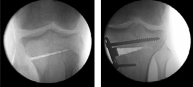
The surgery is most commonly done under a general anaesthetic. The leg alignment is usually changed by cutting the tibia (shin bone) just below the knee, and then opening up the break to allow insertion of a wedge of bone. The size of wedge that is inserted determines the eventual alignment, and is decided from your preoperative X-rays and also X-rays taken during the surgery. Please see the diagram.

X-rays of a tibial osteotomy, showing the opening of the wedge, fixation with a plate and screws, and correction of the alignment.
After creating the gap and checking the alignment, a wedge of bone is inserted to fill the gap. This wedge is either taken from your pelvis, or can be donated or artificial bone. Dr Parker will discuss this with you preoperatively. The position is then held with a plate and screws, a drain inserted, and a brace applied. You will remain in hospital after surgery until you can safely walk with crutches. The physiotherapist in hospital will help you with this. Typically this takes about three to five days.
After Surgery
After discharge from hospital, you will see Dr Parker two weeks later to check the wound. Your brace is worn for 6 weeks, and you use crutches for 12 weeks. During the second 6 weeks you gradually increase the weight on your leg, whilst remaining protected with crutches. An x-ray is taken at six weeks to assess healing of the bone, prior to gradually increasing the weight you put on your leg.
Rehabilitation
Rehabilitation with a physiotherapist is commenced 6 weeks after surgery when healing of the bone is progressing. This is aimed at restoring your movement and muscle strength.
It takes most patients about six months to fully recover from high tibial osteotomy. It is possible to resume a sedentary job three to four weeks after surgery, if this can be done on crutches. It is usually at least 3 to 4 months before physical work is possible, and between 6 – 12 months before sport can be resumed.
Results
Tibial osteotomy usually results in good pain relief and improvement in function. There is no cure for arthritis, and osteotomy does not reverse arthritis but should slow its rapid progression. Osteotomy is typically used in the young or active patient (less than 60), whereas older less active patients would more commonly undergo knee replacement. Knee replacements do not last forever, but in general 90% of knee replacements are still working well after 10 years. We believe that today’s knee replacements are constantly improving but obviously it will take several years to be sure. The wisdom of performing an osteotomy is that it will allow the native knee to survive longer. The older a patient is at the time of knee replacement, the more likely the replacement will last the patient the remainder of their life.
Replacing a knee in a patient who has had a prior tibial osteotomy may be slightly more difficult than performing a primary knee replacement, but is easier and achieves better results than redoing a knee replacement and is usually very successful.
Most patients feel improvement in their knee following tibial osteotomy. A few (5-8%) are unimproved and 2% are worse. The improvement seen following tibial osteotomy lasts a variable time depending on how well the patient cares for the knee, as well as the degree of damage already done by arthritis, and the inherited quality of the articular cartilage in the joint. For over 70% of patients the improvement following osteotomy lasts for 10 years or more.
Complications
Infection: Deep bony infection is very rare but if this occurs and is untreated, serious problems follow.
Blood Clots: Medication and stockings are used to help prevent clots. A clot which travels to the lung can be fatal although this is extremely rare. Chest pain and calf pain can be symptoms of a clot and must be reported to Dr Parker immediately.
Poor Bone Healing: In approximately 2 – 3% of patients, the bone may not fully heal or slip in position whilst healing. This is monitored by x-rays of the bone. Occasionally, revision surgery may be required to promote bone healing.
Nerve and Vessel Injury: Major nerves and arteries which supply the leg are in the vicinity of the surgery. Although rare, damage to these is possible.
Other complications include haematoma, superficial infection and knee stiffness. Please feel free to discuss these with Dr Parker.
Costs of Knee Arthroscopy
Dr Parker’s charges and any associated gap not covered by Medicare and your health fund will be discussed with you when your surgery is arranged. Please feel free to discuss any aspect of this which is unclear to you, either with Dr Parker or his secretary.
If you have any questions concerning your surgery, its risks, benefits, likely outcome or complications please do not hesitate to contact Dr Parker.